

GENERAL HOSPITAL
HANOI - VIENTIANE

 Tiếng Việt
 ຈັນລາວ
 English
Electroencephalogram
CATEGORY

Electroencephalogram
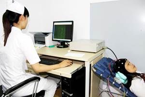
Electroencephalogram records the bioelectrical activity of the brain using electrodes placed on the scalp, in rare cases placed on the surface of the cerebral cortex, or in the brain matter.
These electrical activity manifests as waves on the electroencephalogram.
I. BENEFITS OF EEG SURVEY
It is especially important to detect brain dysfunction in the following neurological diseases:
1. Diagnosis and monitoring of epilepsy or other seizure disorders.
2. Support the diagnosis of brain death.
3. Assess level of awakening during anesthesia.
It also can monitor brain function in other diseases such as:
1. Brain tumors.
2. Head injury.
3. Brain dysfunction.
4. Encephalitis.
5. Stroke.
6. Sleep disorders.
7. Dementia.
II. INTRODUCTION OF ELECTROLOGY
1. Basic types of brain waves
Alpha: frequency 8-13 Hz, amplitude 20-100 microVolt, sinusoidal shape, clearly distributed in the posterior occipital, occipital, and temporal regions.
Beta: frequency >13 Hz, amplitude < 20 microVolt, dominant in the fronto-central region.
Theta: frequency 4-7 Hz, amplitude 30-60 microVolt.
Delta: frequency < 4Hz, amplitude is usually high.
2. Noise waves
Physiological disturbances: electromyography, electrocardiogram, eye movements, tongue movements, skin perspiration, respiration, etc.
Non-physiological: due to electrodes, equipment, environment.
3. EEG normal
In awake adults: dominant posterior rhythm at 8.5-11 Hz, and rapid, low-frequency activity in the anterior leads.
In children: posterior dominant rhythm has lower frequency and higher amplitude, sometimes with posterior slow waves.
Some normal variations: such as Mu rhythm (negative potential, frequency 8-10Hz, lost when moving the opposite hand), slow alpha variation, fast alpha, lamda wave, occipital positive spike wave in sleep (POST- positive occipital sharp transient of sleep),...
4. Common abnormalities on EEG
Slow waves: may be seen in structural brain damage (slow waves are often localized), or are closely related to epileptic activity, or due to encephalopathy
Amplitude abnormalities:
Locally low amplitude due to cortical damage, intracranial or extracranial space-occupying lesions.
Diffuse low amplitude is seen in patients with anxiety causing decreased synchronicity of cortical activity, but can also be seen in cases of cortical damage or cortical dysfunction.
Isoelectric electroencephalogram.
Locally increased amplitude in cranium: high amplitude beta activity, increased sharpness of waves (breach rhythm)
Cyclic activities:
Periodic lateralized epileptiform discharges (PLEDs), sometimes occurring independently in two hemispheres (biPLED). These abnormalities can be seen in acute brain injuries such as stroke, encephalitis, and can also be seen in cases of long-term epilepsy.
Generalized cyclic activity: may be seen in metabolic encephalopathy, poisoning, hypoxia, Creutzfeldt-Jacob disease (mad cow disease).
Burst-suppression pattern or suppression-burst pattern: seen in severe encephalopathy
Seizures: EEG will record whether there are epileptic discharges, focal or general, and where. Seizure-like discharges include: spikes, spikes, spike-wave complexes, spikes-slow waves, multiple spikes, multiple spikes-slow waves, multi spikes-waves, multi-spikes-slow waves.
III. WHAT ARE THE HARMFUL EFFECTS OF EEG MEASUREMENT?
You may experience some discomfort during the measurement as the technician attaches the electrodes to your scalp. Relax because EEG is a safe and harmless method, without any current flowing into your body, EEG only helps to record the electrical activity of your brain.
IV. PROCEDURE OF ELECTROLOGY RECORDING
1. Routine EEG procedure: Lasts about 20 minutes. First, let the patient close his eyes to relax for 3-4 minutes, then perform the eye-opening test (open eyes for 10 seconds, then close the eyes), rest for 5 minutes after the eye-opening test, then perform a deep breathing test (hyperventilation) for 3 minutes, and perform light stimulation afterwards. End the EEG recording by having the patient open/close his eyes one more time.
Eye-opening test (Berger's test): Helps determining the alpha rhythm suppression when opening the eyes and the patient's waking status.
Hyperventilation: This test will be done in 3 minutes. It should not be performed in patients with severe cardiovascular disease, severe cerebrovascular disease, and chronic obstructive pulmonary disease.
Photic stimulation: Starting at a stimulation frequency of 3 cycles/sec, then gradually increasing to 30 cycles/sec. The excitation time at one frequency is 10s and the rest time between the two excitation frequencies is also 10s. This test should be discontinued as soon as the patient develops a photoconvulsive response.
Sleep deprivation: This is a test that helps to increase the ability to detect seizure activity and help record sleep electroencephalograms.
V. WHAT DO YOU NEED TO PREPARE BEFORE MEASUREMENT?
Wash your hair clean the night before the measurement day, do not use hair conditioner, hair gel.
Avoid drinking coffee on the day of EEG measurement.
Make it clear to your doctor if you are taking medication, having any pacemaker inserted, etc.
If you have an appointment for a sleep EEG, you must stay up late, get up early the night before (sleep at 0:00 and wake up at 3 o'clock), do not sleep while waiting for the EEG.